A rounded upper back is not automatically a disease requiring treatment. The important questions are whether the curve is flexible or rigid, whether it is changing, and whether pain, nerve symptoms, breathing limits, or a possible fracture point to an underlying spinal problem. By the end, you will know which signs warrant routine, expedited, or urgent evaluation and what testing a clinician may use.
Key takeaways
- A curve that stays rigid during extension needs clinical assessment.
- Seek urgent care for weakness, numbness, walking problems, or bladder changes.
- Children and teenagers need review for worsening or painful spinal curvature.
- Treatment depends on symptoms, curve cause, progression, and scan findings.
How can you tell flexible postural rounding from structural kyphosis?
A rounded upper back that straightens when you lie down or actively extend your spine is more consistent with flexible postural kyphosis. If a rigid curve remains during extension, arrange assessment for structural kyphosis caused by Scheuermann disease, vertebral fracture, congenital vertebral abnormality, or degenerative change.
| Feature | Flexible postural rounding | Structural kyphosis |
|---|---|---|
| Position | Straightens lying down or extending | Persists during extension |
| Typical cause | Slouching or muscle imbalance | Vertebral shape, fracture, growth-related change, or degeneration |
| Next step | Posture and movement assessment | Clinical examination and possible standing radiographs |
Visual appearance alone cannot establish pathological kyphosis. A clinician considers the measured curve, flexibility, symptoms, age, and cause. On a standing lateral radiograph, thoracic kyphosis commonly falls near 20°–45°; that range provides context, not a diagnosis or automatic treatment threshold.
Arrange an appointment for worsening curvature, persistent pain, a curve newly visible in an adult, loss of height, or difficulty standing, walking, or breathing. A curve that becomes more noticeable during a child’s or teenager’s growth spurt also deserves evaluation, even when pain is mild.
Standing lateral radiographs show the curve under gravity and allow Cobb-angle measurement, while examination determines whether the back actually moves freely.
Do not force extension through pain. A persistent rigid curve needs evaluation because exercises alone will not correct an underlying vertebral problem.
Which symptoms mean you should arrange prompt or urgent evaluation?
Arrange a routine assessment for a persistent or progressively worsening curve, ongoing pain, loss of height, or a newly visible curve in an adult. Do not treat new adult kyphosis as poor posture automatically.
| Situation | Action | Why |
|---|---|---|
| Routine symptoms | Book a clinical appointment | The cause may be osteoporosis, prior fracture, inflammatory disease, infection, tumour, or degenerative spinal change. |
| Expedited evaluation | Arrange prompt review for severe or rapidly worsening back pain, night pain, fever, unexplained weight loss, a history of cancer, significant immune suppression, or a newly developing curve | These kyphosis warning signs can indicate infection, malignancy, or fracture. |
| Urgent symptoms | Seek urgent medical assessment for new leg weakness, numbness, difficulty walking, loss of coordination, or new bowel or bladder dysfunction | These neurological symptoms can signal spinal-cord or nerve-root compression. |
Sudden severe pain after a fall needs prompt assessment for a vertebral compression fracture, especially if you are older or have osteoporosis. A compression fracture can also follow minor trauma or occur without a memorable injury.
Adult kyphosis has different causes, so posture exercises alone are not appropriate until the cause is understood. An examination and, when indicated, imaging can identify fracture or another condition that exercises would not treat.
When should children, teenagers, and older adults be assessed?
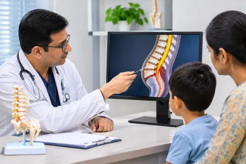
A child or teenager whose roundback increases during a growth spurt should be assessed, even when pain is mild. Progression can occur before skeletal maturity, when remaining growth affects whether observation, therapy, or bracing is useful.
1. Ask for evaluation if the curve is becoming more visible, remains when the child actively straightens, or causes persistent pain. Scheuermann kyphosis is a structural deformity, not ordinary slouching: standing radiographs show wedging of at least 5° in three or more consecutive vertebrae, along with endplate irregularity or Schmorl nodes.
Poor posture is flexible and lacks these structural findings.
2. Arrange assessment for an older adult with a new or worsening curve, loss of height, or new back pain, even without a clear injury. The clinician needs to look for an osteoporotic compression fracture and osteoporosis; fractures can follow minor trauma or occur without a memorable event.
3. Seek earlier review when the curve is rigid, rapidly worsening, or accompanied by weakness, numbness, balance difficulty, or breathing problems. Inflammatory disease, infection, tumour, congenital vertebral abnormalities, and age-related degeneration change the evaluation pathway and may require different imaging or treatment.
Early assessment matters because decisions depend on remaining growth, fracture risk, neurological status, and whether the curve is still flexible. A flexible curve may be observed or treated with exercises, while a progressive structural curve or fracture needs a cause-specific plan.
What happens during kyphosis evaluation, and when are scans needed?
Evaluation starts with a clinical assessment of when the curve and pain began, whether they are progressing, and whether symptoms occur at night. The clinician asks about prior fractures, cancer, inflammatory disease, and effects on work, sleep, walking, standing, horizontal gaze, and breathing.
The examination checks posture, spinal flexibility, height loss, balance, gait, strength, sensation, reflexes, and bowel or bladder symptoms.
Imaging is selected according to those findings:
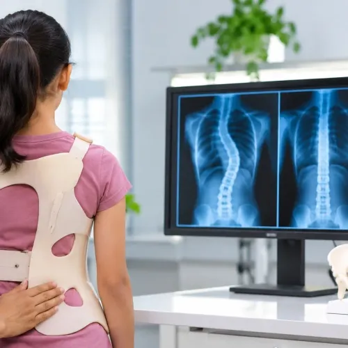
- A standing lateral spine radiograph shows how gravity affects the curve and permits measurement of the sagittal Cobb angle. Standing front and side views of the whole spine assess scoliosis and overall alignment.
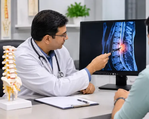
- MRI for kyphosis is considered when there are neurological findings, suspected spinal-cord compression, disproportionate or rapidly worsening pain, infection, tumour, an atypical curve, or a need to assess the cord and discs before surgery.
- CT provides detail about fractures, vertebral bone, congenital anatomy, and operative planning. It is not usually needed for uncomplicated, flexible postural rounding without red flags.
Images cannot establish the cause alone; interpret them alongside symptoms and examination findings. A new weakness, numbness, walking difficulty, poor coordination, or bowel or bladder change needs urgent assessment rather than routine scanning.
When does evaluation lead to treatment rather than observation?
Evaluation does not automatically mean surgery. Mild, flexible, or minimally symptomatic curves may be managed with:
- Observation and repeat examination or imaging when progression is a concern
- Activity guidance that avoids aggravating pain without unnecessary restriction
- Physical therapy for posture, spinal mobility, strength, and function
- Treatment of the underlying cause, such as osteoporosis or an inflammatory disorder
For a growing adolescent with structural Scheuermann kyphosis, bracing is mainly considered while growth remains. Its ability to remodel the curve declines substantially after skeletal maturity, so delaying assessment can narrow this option.
Surgery enters the kyphosis treatment discussion when one or more of these findings is present:
- Substantial or progressive deformity
- Disabling pain despite appropriate nonoperative care
- Major sagittal imbalance affecting standing or horizontal gaze
- Neurological compromise from cord or nerve compression
- A correctable structural cause
Appearance alone, or crossing one Cobb-angle value, is not enough. In adults with degenerative kyphosis, the decision also weighs pain-related disability, inability to maintain horizontal gaze or standing balance, nerve compression, and response to conservative care. Major reconstruction carries kyphosis surgery risks, including blood loss, nonunion, implant failure, and junctional deformity.
A neurosurgical consultation with Dr Amit Aiwale-Best Neurosurgeon in Thane can connect examination and imaging findings to an appropriate plan when symptoms, fracture risk, or neurological signs make specialist assessment useful.
Related service
Spine Kyphosis Spine Kyphosis is an exaggerated, abnormal forward curve of the upper back (thoracic spine), leading to a “hunchback” or slouching posture. View service → |
Frequently asked questions
How can you tell flexible postural rounding from structural kyphosis?
Flexible postural rounding straightens when you lie down or actively extend your spine. A rigid curve that remains during extension needs assessment for structural kyphosis.
Which symptoms mean you should arrange prompt or urgent evaluation?
Arrange prompt evaluation for persistent pain, a worsening curve, or unexplained height loss. Seek urgent care for new weakness, numbness, walking difficulty, or bladder or bowel changes.
When should children, teenagers, and older adults be assessed?
Assess children and teenagers when the curve progresses, causes pain, or remains rigid. Older adults need review after a fall, sudden back pain, height loss, or suspected vertebral fracture.
What happens during kyphosis evaluation, and when are scans needed?
Evaluation includes medical history, posture and spine examination, movement testing, and a neurological check. X-rays assess alignment and vertebral shape; MRI or CT is added for neurological symptoms, suspected fracture, or unclear findings.
When does evaluation lead to treatment rather than observation?
Treatment is considered when kyphosis causes pain, neurological problems, functional limits, progression, or an underlying condition requiring care. Mild, flexible, symptom-free curves may be monitored with exercise and follow-up.